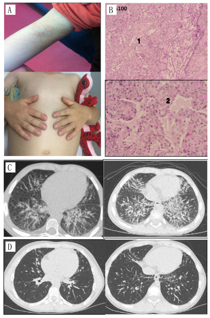
Figure 1.
(A) specific rash and nail clubbing; (B) Lung biopsy, 1: parenchymal crystals, 2: foamy histiocytes; (C) CT, diffuse interstitial syndrome, predominantly in the two lower lobes with inter-lobular septa, peribronchovascular thickening and some peribronchovascular and subpleural micronodules. Increased diffuse interstitial syndrome, more extensive in the upper lobes and the anterior territories of the lower and middle lobes; (D) CT two years after introduction of baricitinib: significant improvement in bilateral interstitial lung disease.

