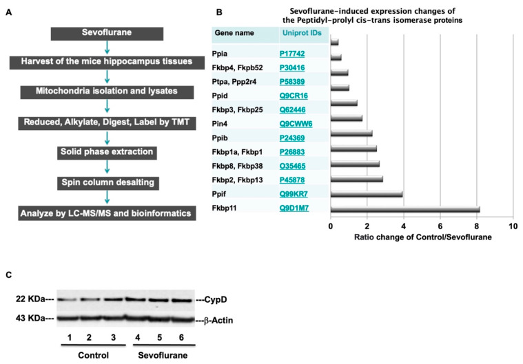
Figure 5.
Proteomics analyses identify mitochondrial proteins in the hippocampus of mice. (A) Experimental design and workflow for quantitative profiling of mice hippocampus mitochondrial proteomics after the sevoflurane anesthesia. (B) Quantitative proteomic analysis of the abundance ratio of Peptidyl-prolyl cis-trans isomerase (Ppif) associated proteins after the sevoflurane anesthesia. (C) Sevoflurane treatment (lanes 4 to 6) discernibly increases the levels of CypD as compared to the control condition (lanes 1 to 3) in the hippocampus of WT mice; there is no significant difference in β-actin levels between the groups.

