Figure 6.
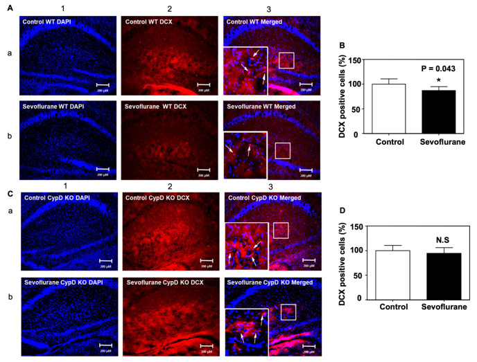
CypD KO mitigates the sevoflurane-induced decrease of DCX-positive cells in the hippocampus of mice. (A) Representative images of immunofluorescent staining DCX in the hippocampus of WT mice. Column 1 is the DAPI (blue) nuclear staining, column 2 is the DCX (red) staining, and column 3 is the merged staining image. Sevoflurane (row b) decreases the number of DCX-positive cells compared to the control condition (row a) in the hippocampus of WT mice. (B) Quantification of the image shows that sevoflurane (black bar) significantly decreases the number of DCX-positive cells compared to the control condition (white bar) (* p = 0.043). (C) Representative images of immunofluorescent staining DCX in the hippocampus of CypD KO mice. (D) Quantification of the image shows that the sevoflurane anesthesia (black bar) does not change the number of DCX-positive cells in the hippocampus of CypD KO young mice compared to the control condition (white bar). N = 5–6 in each group. Student’s t-test was used to analyze the data. The arrows indicate the DCX positive cells in hippocampus. The Box indicates the zoom in image area in staining. N.S: non-significant differences.

