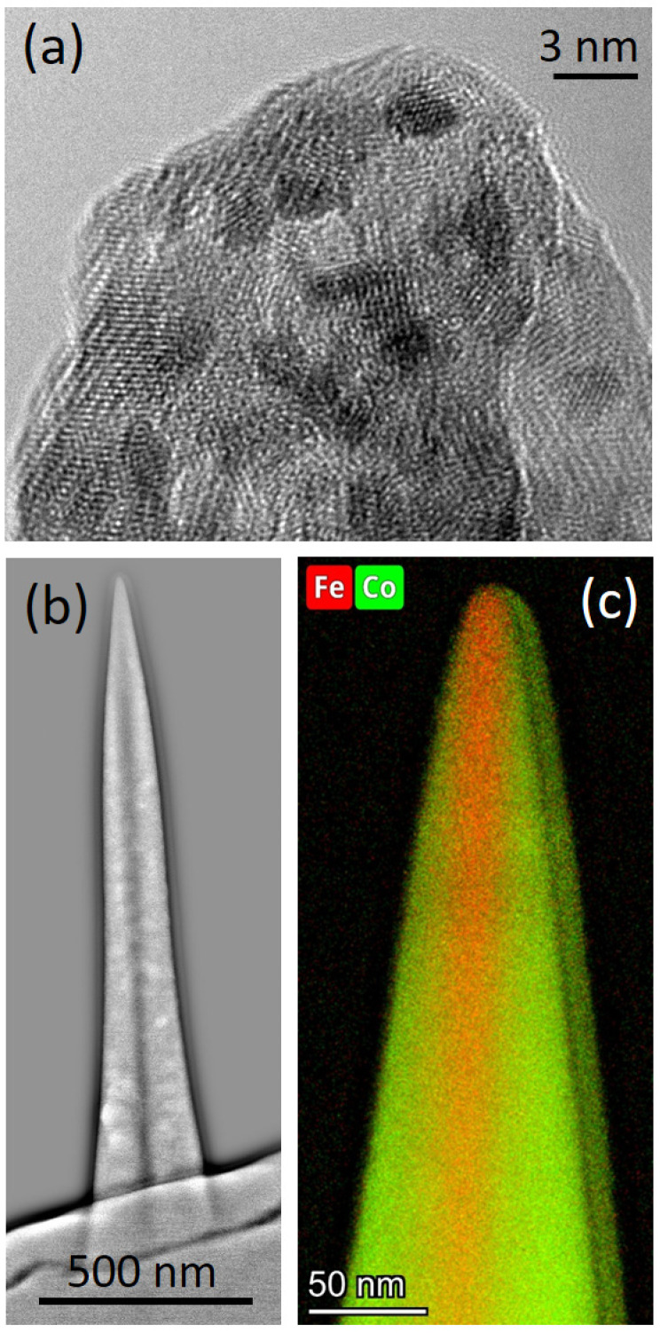
Figure 5.
TEM characterization of Co3Fe pillars. (a) high-resolution TEM image of the tip region confirms the high degree of crystallinity, the contamination-free character, and the sharp apex. (b) high-pass filtered TEM-HAADF image of a Co3Fe α-pillar, which reveals a dark core in the center. (c) Scanning-TEM EDX map of Fe and Co distribution at the tip region, which reveals Fe-richer core, while surrounding areas are closer to the expected Co3Fe composition.

