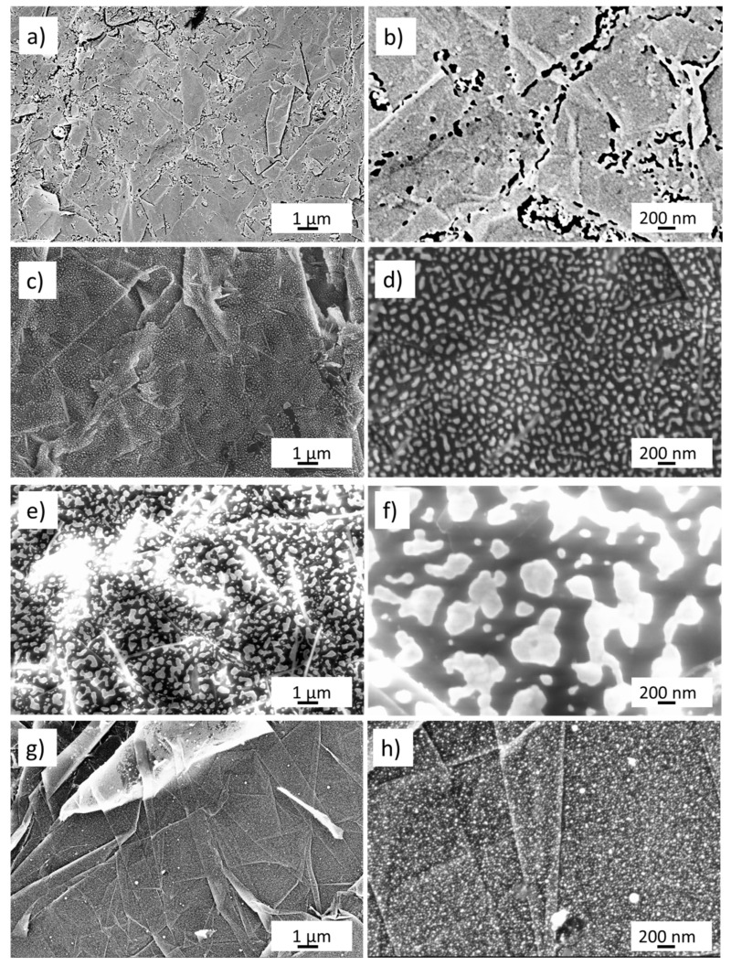
Figure 1.
Field emission scanning electron microscopy pictures of (a,b) gold nanoporous obtained by five cycles of scanning potential between −0.5 and 1 V in NaOH 0.1 M; (c,d) gold layer 8 nm thin dewetted at 300 °C; (e,f) gold layer 17 nm dewetted at 400 °C; (g,h) gold layer 17 nm dewetted by laser at 0.5 J cm−2 fluence.

