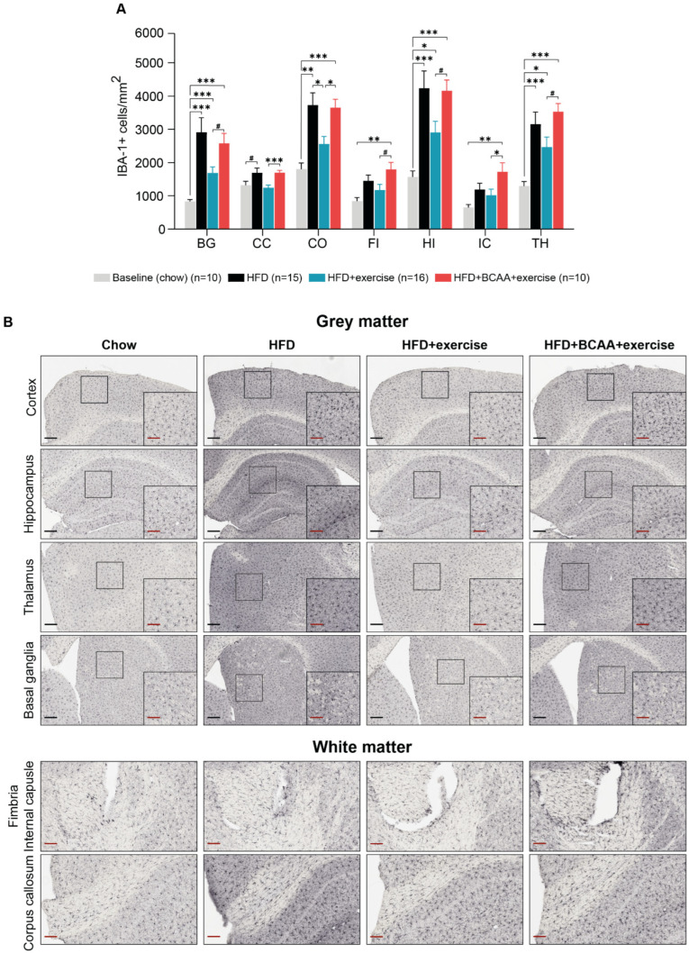
Figure 11.
Neuroinflammation. Immunohistochemical analysis of ionized calcium-binding adapter molecule (IBA-1), as a measure of neuroinflammation in different brain regions of interest (basal ganglia (BG), corpus callosum (CC), cortex (C), fimbria (FI), hippocampus (HI), internal capsule (IC), and thalamus (TH)). (A) The number of IBA-1+ cells/mm2 was analyzed in young chow-fed animals (baseline), HFD, HFD + exercise, and HFD + BCAA + exercise groups. (B) Representative images of the IBA-1 staining (black scale bar = 200 µm; red scale bar = 100 µm). High-fat diet (HFD), branched-chain amino acids (BCAA). Data are presented as mean ± SEM. * p < 0.05, ** p < 0.01, *** p < 0.001, # 0.05.

