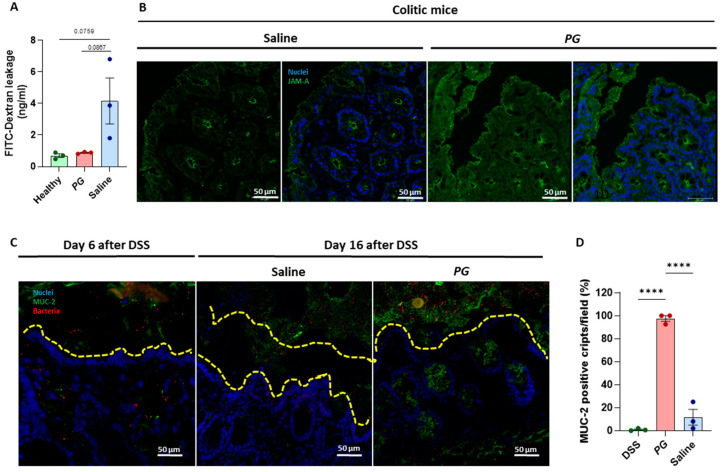
Figure 2.
PG restored the intestinal barrier in vivo with the formation of a mucus layer. (A) Intestinal permeability was evaluated through FITC-dextran assay in healthy and PG or saline-treated mice, quantifying FITC-dextran levels in murine sera. (B) Representative images of immunofluorescence staining of JAM-A (green) in the epithelial layer and crypts of colitic mice during the recovery phase subjected to saline or PG. (C) Fluorescence in situ hybridization for bacterial rRNA (red) and immunofluorescence staining of MUC-2 (green); and (D) quantification of MUC-2 positive cripts for field (40X)was performed in the colonic mucosa of colitic mice before and after the recovery phase with the administration of saline or PG. (DAPI = nuclei in blue. Scale bar = 50 µm. Data are presented as mean ± SEM. One-way ANOVA test. **** p < 0.0001. n = 3 mice/group.

