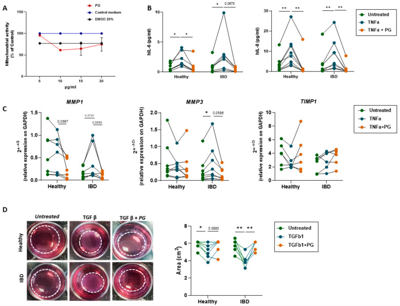
Figure 6.
PG reverted fibroblast activation, decreasing inflammatory cytokines, MMP expression, and the contractile phenotype. (A) MTT assay was performed on primary intestinal fibroblasts isolated from noninflamed and inflamed areas of IBD patients after stimulation with PG at different concentration (5, 10, 15, 30 µg/mL) for 48h. Primary fibroblasts untreated or stimulated with 20% DMSO were used as control. (B) IL-6 and IL-8 levels (PG/mL) in healthy and IBD fibroblasts treated for 48h ± TNF-α or TNF-α + PG. (C) Quantitative Real-Time PCR analysis of MMP1, MMP3, and TIMP1 mRNA expression in healthy and IBD fibroblasts stimulated ± TNF-α or TNF-α + PG. Gene expression was normalized to GAPDH. (D) Representative images of contraction assay performed on healthy and IBD fibroblasts stimulated ± TGF-β or TGF-β + PG. The collagen circles are highlighted by the dashed white line. The circle area (cm2) was measured in healthy and IBD fibroblasts stimulated ± TNF-α or TNF-α + PG. Data are presented as mean ± SEM. Paired t-test * p < 0.05, ** p < 0.01. n = 8 healthy/group, n = 8 IBD (5 CD and 3 UC)/group.

