Figure 7.
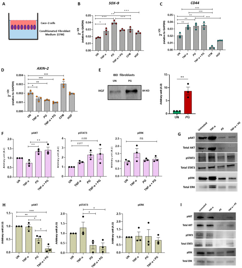
PG promoted intestinal epithelium repair by regulating fibroblast activity. (A) Experimental design of Caco−2 cells cultured for 24 h with conditioned medium (CFM) derived from IBD fibroblasts stimulated ± TNF-α, PG, or TNF-α + PG. (B−D) Quantitative Real-Time PCR analysis of SOX−9, CD44, and AXIN−2 mRNA expression in Caco-2 cells treated ± TNF-α, PG, TNF-α + PG, CFM derived from fibroblasts stimulated with PG or HGF. Gene expression was normalized to GAPDH. (E) Western blot analysis of HGF in the supernatants of IBD fibroblasts ± PG. HGF was normalized on total proteins (F,G) Western blot analysis of pAKT, Total AKT, pSTAT3, Total STAT3, pERK, and Total ERK in Caco−2 cells ± TNF-α, PG or TNF-α + PG. (H,I) Western blot analysis of pAKT, Total AKT, pSTAT3, Total STAT3, pERK, and Total ERK in IBD fibroblasts ± TNF-α, PG or TNF-α + PG. Phosphorylated AKT, STAT3 and ERK were normalized on total AKT, STAT3, and ERK, respectively. Data are presented as mean ± SEM. One-way ANOVA test. * p < 0.05, ** p < 0.01, *** p < 0.001. n = 3/group.

