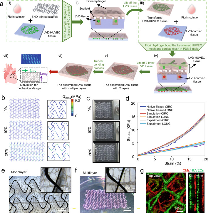
Fig. 6. Engineering of 3D pre-vascularized LVD-cardiac tissue constructs with the programmed mechanical property.
a The transfer and assembly process of the multiple LVD tissues. FEA simulations (b) and macroscopic images (c) of the 3D tissue composite upon uniaxial stretching (from top to bottom: 0%, 10%, and 20%) along the CIRC direction. d The stress–strain curves along the CIRC and LONG direction among the corresponding FEA calculations, experimental measurement, and adult rat right ventricular myocardium. n = 3 independent samples. e Bright-field of the transferred monolayer LVD-rat-cardiac tissue. The macroscopic (f) and confocal (g) images of the assembled 4-layer LVD-rat-cardiac construct, consisting of red LVD-rat-cardiac and green LVD-HUVEC tissues superimposed with each other.

