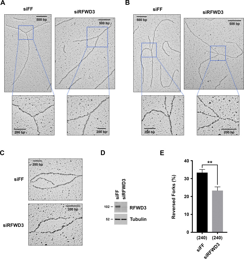
Figure 5.
RFWD3 promotes the reversal of stalled replication forks. (A and B) Representative images of normal (A) and reversed (B) replication forks detected by electron microscopy (EM) in control or RFWD3-depleted U2OS cells upon HU treatment. (C) Representative images of DNA replication bubbles detected by EM in HU-treated control and RFWD3-depleted U2OS cells. (D) Detection of RFWD3 levels in U2OS cells transfected with siRFWD3-4 for the experiment in Fig. 5 E. See Fig. S3 C for the depletion efficiency of other replicates used in Fig. 5 E. (E) Percentage of reversed replication forks detected by EM in control and RFWD3-depleted U2OS cells upon HU treatment (2 mM for 5 h). The mean and SEM from three independent experiments are shown (**P < 0.01, t test, see Fig. S3 C). The number of replication intermediates analyzed for each condition is indicated in parentheses.

