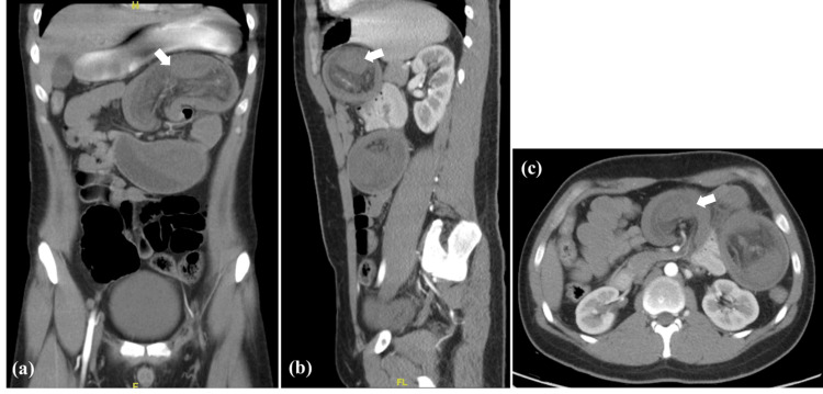
Figure 1. CT scan with oral and IV contrast: (a) coronal view, (b) sagittal view, (c) axial view, all showing dilated proximal jejunal loops (arrows) at the left upper abdomen with configuration of bowel within bowel, indicating the possibility of jejunojejunal intussusception.
Congestion of the adjacent mesentery is also present with no passage of oral contrast beyond the intussusception point.

