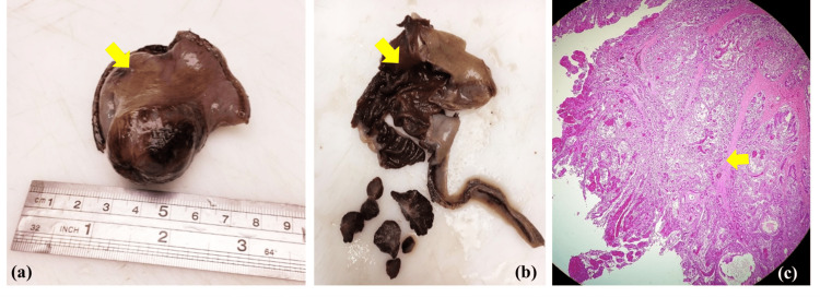
Figure 3. (a) Gross inspection of the resected specimen shows a small intestinal segment (arrow) measuring 6 cm × 4 cm × 4 cm with stapled ends, polypoid intussusception, and gangrenous serosa. (b) Cut surface reveals a 3 cm × 2.5 cm pedunculated polyp (arrow) with hemorrhagic surface and surrounding gangrenous mucosa. (c) The histological view (hematoxylin and eosin, ×100) shows a pedunculated polyp lined by normal glands and crypts of small intestine with goblet cells.
There is haphazard arrangement of gland clusters with treelike branching in addition to smooth muscle bundles and fibrosis within the core of the polyp (arrow). The specimen is negative for malignancy.

