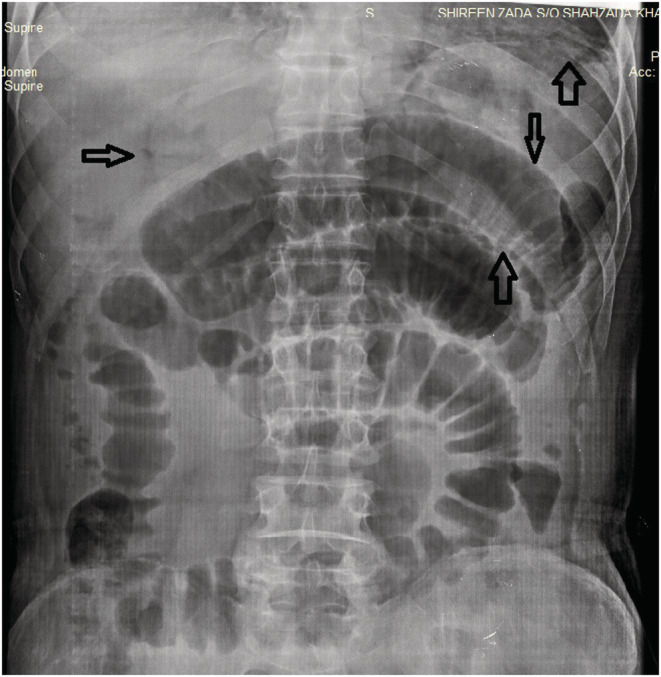
FIGURE 1.

X‐rays of the supine abdomen showing dilated bowel loops, free air, air in the portal vein and Rigler's sign (sign of pneumoperitoneum seen when gas is outlining both sides of the bowel wall). No abnormal calcification was found. Normal densities of soft tissue were identified. No radio‐opaque stone was seen in the KUB area.
