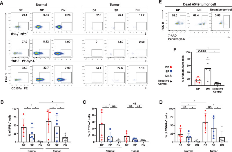
Figure 3.

Functions of the trNK cells in NSCLC. (A−D) The percentages of IFN‐γ+ (B), TNF‐α+ (C), and CD107a+ cells (D) in DP, SP, and DN NK cells from the normal and tumor tissues. Data are shown as the representative flow analysis (A) and mean ± SEM (B−D). (E−F) The percentages of the dead A549 lung cancer cells after co‐culture with sorted DP and DN NK cells. Negative control refers to as the A549 cells cultured alone. Data are shown as the representative flow analysis (E) and mean ± SEM (F). Data were obtained from the normal and paired tumor tissues resected from ten of 52 operative NSCLC patients, pooled from three independent experiments. Symbols represent one individual sample. *p < 0.05, **p < 0.01, ***p < 0.001 (two‐tailed t‐test and Wilcoxon signed rank test in B−D and F).
