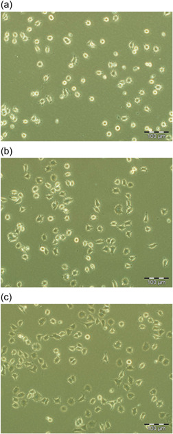
Figure 2.

Cell morphology in different concentrations of collagen IV. NS‐1 cells were cultured in Dulbecco's modified eagle's medium (DMEM) media with 15% horse serum and 2.5% foetal bovine serum in a T25 flask as described in the Section 2. Cells were seeded into six‐well plate with each well coated with 3 µg/ml (a), 10 µg/ml (b) or 30 µg/ml (c) collagen IV respectively. After 24 h of incubation, representative phase contrast photomicrographs were taken. (a) Show a majority of rounded, loosely attached phase‐bright cells. (b) Shows a majority of spread out, firmly attached phase‐dark cells. (c) Shows almost all the cells are spread out and strongly attached. Scale bar (a, b, c) 100 µm.
