FIGURE 2.

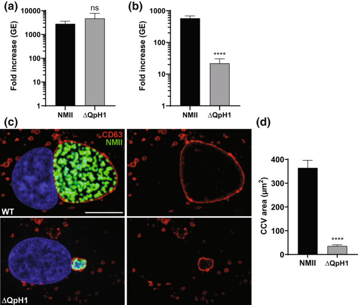
Coxiella burnetii ΔQpH1 has a severe intracellular growth defect. Replication of wild‐type NMII C. burnetii and the ΔQpH1 mutant in (a) ACCM‐D and (b) Vero cells. Replication is shown as fold increase in C. burnetii genome equivalents (GE) in axenic media (7 days) and Vero cells (6 days). Results are expressed as the means of results from two biological replicates from three independent experiments. Error bars indicate the standard deviations from the means, and asterisks indicate a statistically significant difference (Student's t test, ****p < .0001) compared with values for wild‐type C. burnetii. ns indicated no significant difference. (c) Confocal fluorescence micrographs of Vero cells infected for 3 days with NMII and ΔQpH1 mutant. CD63 (red) and C. burnetii (green) were stained by indirect immunofluorescence, and DNA (blue) was stained with Hoescht 33,342. Bar, 10 μm. (d) The histogram depicts the mean intensity of CCV area +/− SD of >50 cells for at least three independent experiments. Statistical difference was determined using the Student's t test (****p < .0001)
