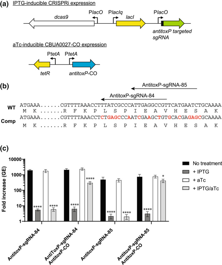
FIGURE 6.

Complementation of antitoxP CRISPRi repression allows restoration of intracellular growth. (a) Schematics of the IPTG‐inducible antitoxP CRISPRi repression system and aTc‐inducible expression of the codon‐optimized antitoxP gene. In the presence of IPTG expression of dCas9‐3x‐c‐myc and antitoxP‐targeted sgRNA occurs from the Plac promoters. The presence of aTc expression of the codon‐optimized antitoxP gene occurs from the PtetA promoter. (b) Partial sequences of the wild‐type (WT) and codon‐optimized antitoxP gene are depicted. The location of the two antitoxP sgRNA targeting sequences is shown with arrows. Nucleotides that were changed are depicted in red. For toxP‐sgRNA‐84, 9 bp of the target sequence was changed and for toxP‐sgRNA‐85, 8 bp of the target sequence was changed. These changes did not affect the protein sequence. (c) Expression of antitoxP‐CO allows complementation of THP‐1 macrophage growth in antitoxP CRISPRi repression strains. Replication is shown as a fold increase in Coxiella burnetii genome equivalents (GE) of antitoxP CRISPRi strains with or without the antitoxP‐CO complement plasmid (pJB‐lysCA‐TetRA‐antitoxP‐CO) after a 5‐day infection of THP‐1 macrophages in the absence of any inducer (no treatment; CRISPRi off/complement off), presence of IPTG (CRISPRi on/complement off), presence of aTc (CRISPRi off/complement on), or presence of both IPTG and aTc (CRISPRi on/complement on). Results are expressed as the means of results from two biological replicates from three independent experiments. Error bars indicate the standard deviations from the means, and asterisks indicate a statistically significant difference (one‐way ANOVA, *p < .05, ****p < .0001) compared with values for the no‐treatment samples
