
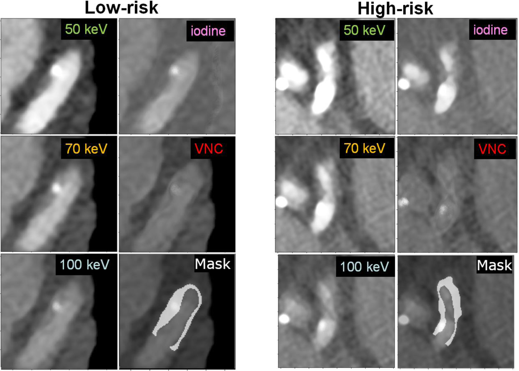
Figure 2.
Sample images (window width/window level: 900/150 HU) of two plaques from two different patients, one classified as low-risk (left) and the other high-risk (right), with their associated segmentation mask overlaid on the 100 keV VMI.
Official websites use .gov
A
.gov website belongs to an official
government organization in the United States.
Secure .gov websites use HTTPS
A lock (
) or https:// means you've safely
connected to the .gov website. Share sensitive
information only on official, secure websites.
Sample images (window width/window level: 900/150 HU) of two plaques from two different patients, one classified as low-risk (left) and the other high-risk (right), with their associated segmentation mask overlaid on the 100 keV VMI.