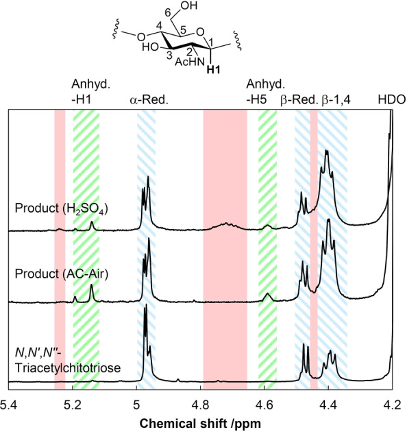
Figure 3.

1H NMR spectra of hydrolysates and standard trimer in D2O‐DMSO‐d6 (1 : 3) mixture. Blue diagonal line: H1 for linear chitin‐oligosaccharides including a small amount of NAG (α‐reducing end, β‐reducing end, β‐1,4‐glycosidic bond); green diagonal line: H1 and H5 for anhydride forms of reducing ends in any oligosaccharide; red fill: H1 for other oligosaccharides with unidentified structures. The chemical shifts for anhydrides were determined using 1,6‐anhydro‐β‐N‐acetylglucosamine.
