
Fig. 7:
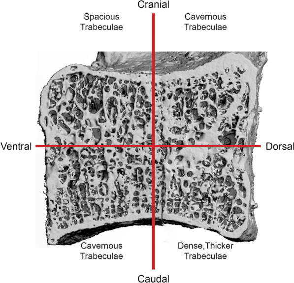
3D µCT reconstruction of a sagittal cut C4 vertebra split into four quadrants corresponding to the cranial–caudal and ventral–dorsal axis. Region-specific descriptions of the trabecular substructure are detailed in the respective quadrant
Official websites use .gov
A
.gov website belongs to an official
government organization in the United States.
Secure .gov websites use HTTPS
A lock (
) or https:// means you've safely
connected to the .gov website. Share sensitive
information only on official, secure websites.
3D µCT reconstruction of a sagittal cut C4 vertebra split into four quadrants corresponding to the cranial–caudal and ventral–dorsal axis. Region-specific descriptions of the trabecular substructure are detailed in the respective quadrant