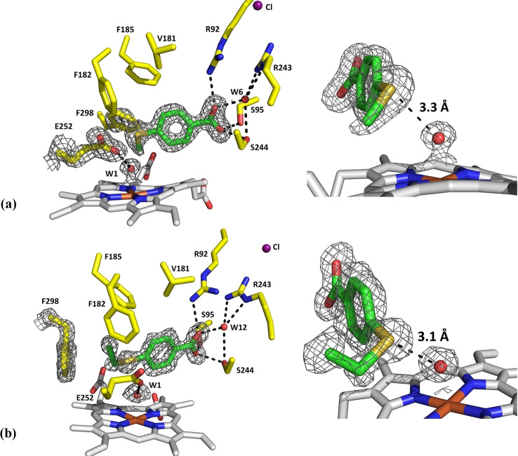
Figure 5.
Crystal structures of the T252E mutant of CYP199A4 in complex with 4‐methylthiobenzoic acid (a; PDB code: 7TP6) and 4‐ethylthiobenzoic acid (b; PDB code: 7TP5). A 2mFo‐DFc composite omit map or feature‐enhanced map of the substrate, the heme aqua ligand (W1), the E252 side‐chain and the phenylalanine 298 residue is shown as gray mesh contoured at 1.5 or 1.0 σ (1.5 Å carve). The sulfur of each substrate interacts with the iron‐bound oxygen ligand (3.3 Å and 3.1 Å, respectively).

