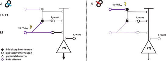
Figure 7. Model of the possible neural circuits involved in the plasticity changes after the PMv‐to‐M1 cc‐PAS.

The large pyramidal neuron (PN) in L5 of M1, which projects to the spinal cord, receives both excitatory (white circle) and inhibitory (black circle) synaptic inputs responsible for the I2‐waves. The PMv projections (violet circle) contact the interneurons both in L2–3 and in L5 of M1 (Ghosh & Porter, 1988). The lightning bolt represents the preferential activation layers of the cc‐PAS stimulation while the shaded circuits indicate the not preferential action sites of cc‐PAS and the thickness of neurons indicates the increase or decrease in their activity. A, the PMv projection that synapses with the interneurons in the deepest layer (L5), preferentially enhanced by the cc‐PAS in PA direction, excites the PN, leading to an increment of the CSE, and inhibits the circuit responsible for the I2‐waves. B, on the other side, the more superficial interneurons populations in L2‐3, probably responsible for the I2‐wave and preferentially activated by the AP stimulation, can inhibit the dendritic arbour of the PN (Jiang et al., 2013), leading to reductions of the CSE, and in parallel enhance the I2‐waves exciting the responsible circuitry.
