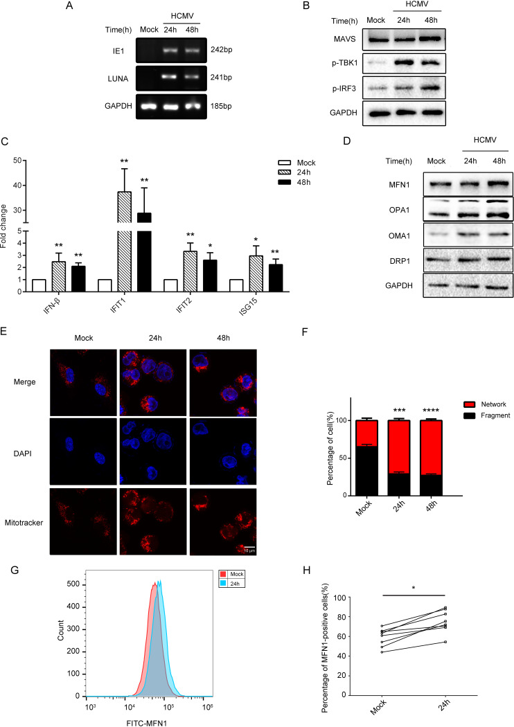
FIG 1.
HCMV infection caused the imbalance of mitochondrial fusion division and induced the expression of MFN1 in THP-1 cells and human monocytes. (A) The levels of HCMV transcripts (IE1 and LUNA mRNA) analyzed by RT-PCR. (B) Western blot of MAVS, p-TBK1, p-IRF3 in lysates from control, and THP-1 cells infected with HCMV (MOI = 5) for 24 or 48 h. GAPDH was used as a loading control. (C) The levels of HCMV transcripts (IFN-β, IFIT1, IFIT2 and ISG15 mRNA) analyzed by RT-PCR. (D) Western blot of MFN1, OPA1, OMA1, and DRP1 in lysates from control and THP-1 cells infected with HCMV (MOI = 5) for 24 or 48 h. GAPDH was used as a loading control. (E) THP-1 cells were infected with HCMV (MOI = 5) for 24 or 48 h, stained, and subjected to confocal microscopy. Uninfected cells serve as a mock control. (F) Mitochondrial perimeter measurements were obtained from multiple independent images and graphed as a percentage of the total number of mitochondria counted for each sample. (G, H) Flow cytometry analysis of the intracellular MFN1 protein expression from donors’ peripheral blood mononuclear cells infected with HCMV (MOI = 5) for 24 h. The data are shown as the means ± standard error of the mean (SEM) of three independent experiments. DAPI, 4′,6-diamidino-2-phenylindole; FITC, fluorescein isothiocyanate; GAPDH, glyceraldehyde-3-phosphate dehydrogenase; HCMV, human cytomegalovirus; IE, immediate early gene; IFIT, interferon-induced protein with tetratricopeptide repeat; IFN, interferon; ISG, IFN-stimulated gene; LUNA, latency unique nuclear antigen; MAVS, mitochondrial antiviral signaling protein; NS, not significant; P > 0.05; *, P < 0.05; **, P < 0.01; ***, P < 0.001.

