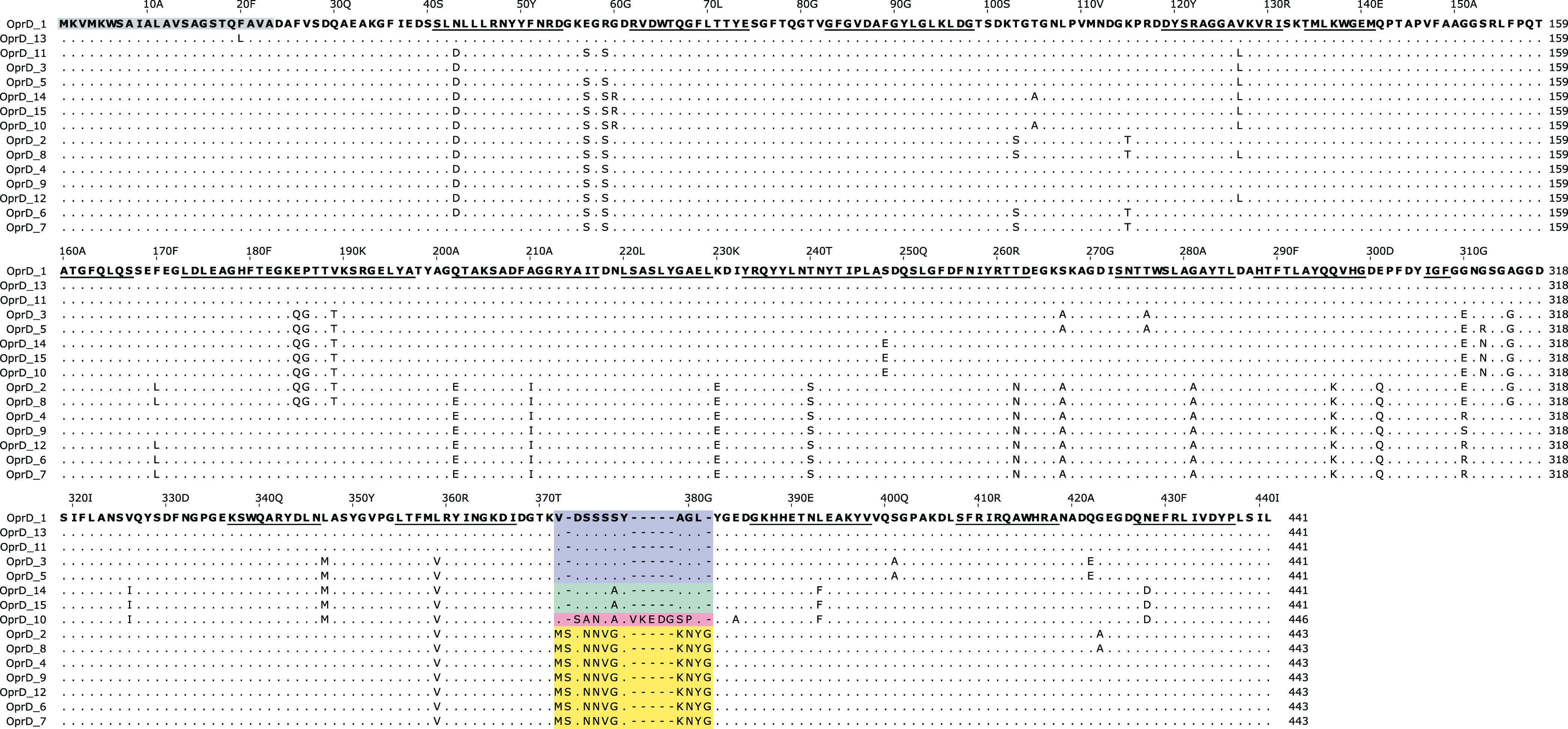
FIG 2.

Alignment of P. aeruginosa OprD variants. The variable region within loop 8 is colored (L810, blue; L810b, green; L812, yellow; L815, red). Scale numbers above the alignment refer to amino acid positions in variant OprD_1 and include the signal peptide (23 amino acids, gray box). β-Strands (according to PDB structure 3SY7) are underlined.
