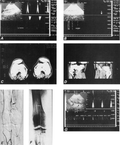
Fig. 3 Case 3: A) Duplex sonogram waveform from a normal left popliteal artery demonstrates a rapid early rise in systole. B) Duplex sonogram from right popliteal artery shows complete occlusion (vertical arrow) and no Doppler signal. C) Dynamic computed tomographic scan with intravenous contrast enhancement demonstrates thrombosis of the right popliteal artery (black arrow), in contrast with patent left popliteal artery (white arrow). Accessory head of the right gastrocnemius muscle (white arrowhead) is clearly demonstrated, in contrast with normal anatomic relationships seen in left limb. D) Phase contrast computed tomographic images in sagittal plane depict increased mass of the 3rd head of the gastrocnemius muscle in the right eg (arrowheads). E) Digital subtraction arteriogram demonstrates a complete segmental occlusion of the right popliteal artery (arrow). Note the absence of atherosclerotic signs in the collateral arteries. F) The length of the occluded segment was 120.6 mm (white line). G) Control postoperative Doppler ultrasonogram demonstrates patent saphenous vein in situ bypass (arrowheads) between the proximal and distal portions of the popliteal artery.

An official website of the United States government
Here's how you know
Official websites use .gov
A
.gov website belongs to an official
government organization in the United States.
Secure .gov websites use HTTPS
A lock (
) or https:// means you've safely
connected to the .gov website. Share sensitive
information only on official, secure websites.
