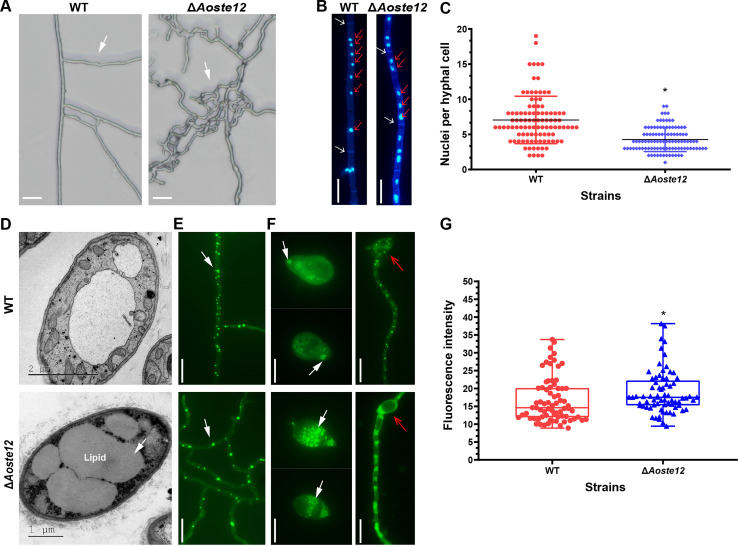
FIG 4.
Comparison of hyphal polar growth, and nucleus and lipid droplet accumulation of wild-type (WT) and ΔAoste12 mutant strains. (A) Comparison of hyphal polar growth of WT and ΔAoste12 mutant. (B) Hyphae were stained with DAPI, and the nuclei of the WT and the ΔAoste12 mutant were observed. White arrow, hyphal septum; red arrow, nucleus. (C) Comparison of cell nuclei in hyphae of the WT and ΔAoste12 mutant. (D) Observation of cellular ultrastructure in hyphae of WT and ΔAoste12 mutant via TEM. (E) Observation of lipid droplets in hyphae of WT and ΔAoste12 mutant by staining with boron dipyrromethene dye. (F) Observation of lipid droplets in conidia and germinated hyphae of spores in WT and ΔAoste12 mutant. White arrow (D to F), lipid droplets. Red arrow, germinated conidium. (G) Comparison of lipid droplet content of WT and ΔAoste12 mutant, which were detected as fluorescence intensity, and determined for at least 70 fields observed under fluorescence electron microscopy. An asterisk indicates a significant difference between the mutant and the WT strain (Tukey’s HSD, P < 0.05). White arrow (D, E, and F), lipid droplet. Scale bar (A, B, E, and F), 10 μm.

