
FIG 2.
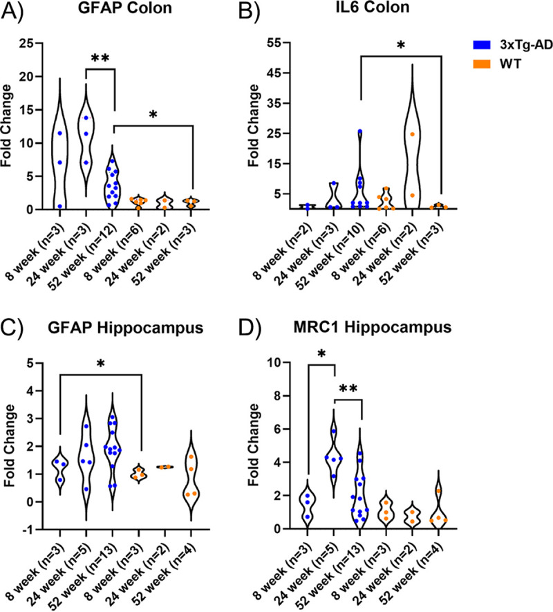
Relative levels of gene expression of GFAP and IL-6 in the colon and GFAP and MRC1 in the hippocampus. The hippocampus and colon from 3xTg-AD and WT mice were collected at 8, 24, and 52 weeks. (A) Gene expression of GFAP (astrogliosis marker) was significantly increased at 24 weeks in 3xTg-AD mice compared to 52 weeks in 3xTg-AD mice (P = 0.009, Mann-Whitney test) and was increased at 52 weeks in 3xTg-AD mice compared to 52 weeks in WT mice (P = 0.0484, Mann-Whitney test). (B) Gene expression of IL-6 was significantly increased at 52 weeks in 3xTg-AD mice compared to 52 weeks in WT mice (P = 0.015, Mann-Whitney test). (C) Gene expression of GFAP (astrogliosis marker) was significantly increased at 52 weeks in 3xTg-AD mice compared to 52 weeks in WT mice (P = 0.049, Mann-Whitney test). (D) Gene expression of Mrc1 (microgliosis marker) was significantly increased at 24 weeks in 3xTg-AD mice compared to 52 weeks (P= 0.004, Mann-Whitney test) and 8 weeks (P= 0.0357, Mann-Whitney test) in 3xTg-AD mice. *, P < 0.05; **, P < 0.01.