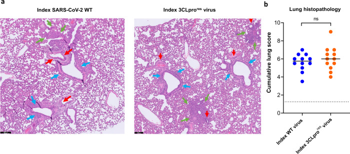
Fig. 2. Histopathology of lungs of Syrian hamsters infected with either the wild-type SARS-CoV-2 or the 3CLpro (L50F-E166A-L167F) resistant virus.
a Representative H&E images of lungs of hamsters infected with 104 TCID50 of either the wild-type (WT) SARS-CoV-2 virus (USA-WA1/2020) or the 3CLpro (L50F-E166A-L167F) nirmatrelvir resistant (3CLprores) virus at day 4 post-infection (pi) showing peribronchial inflammation (blue arrows), peri-vascular inflammation with vasculitis (red arrows), and foci of bronchopneumonia (green arrows). Scale bars, 200 μm. b Cumulative severity score from H&E stained slides of lungs from hamsters infected with the WT virus or the (3CLprores) virus at day 4 pi. Individual data and median values are presented and the dotted line represents the median score of untreated non-infected hamsters. Data were analyzed with the two-sided Mann−Whitney U-test, ns = non-significant (p = 0.5). Data presented are from 2-independent studies with a total n = 12 per group.

