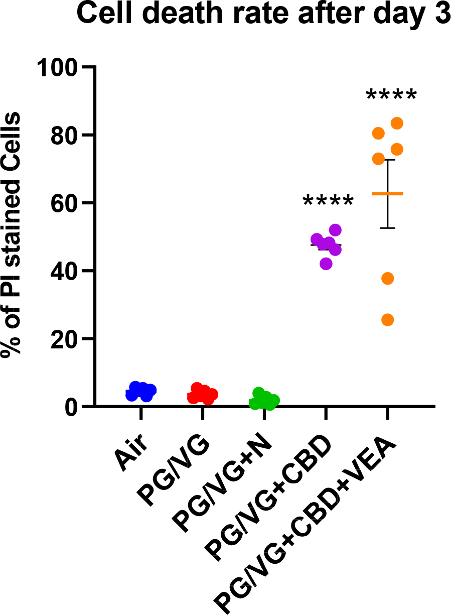
Figure 4: CBD oil- and VEA aerosol-exposed HBECs show high percentages of dead cells after 3 consecutive exposures.

HBECs exposed to aerosols containing CBD oil showed ~45% and CBD oil+VEA ~60% dead cells after the 3rd exposure. Plotted are percentages of propidium iodide (PI)-positive cells, indicating cell death rates for each exposure group. HBE cultures from 6 individual donors (n=6) per exposure group were analyzed in triplicate fluorescence analyses. Mean and SEM values are indicated by major and minor horizontal bars, respectively. Statistical significance was determined by Dunn’s multiple comparison test; the p-value is indicated by **** ≤ 0.001.
