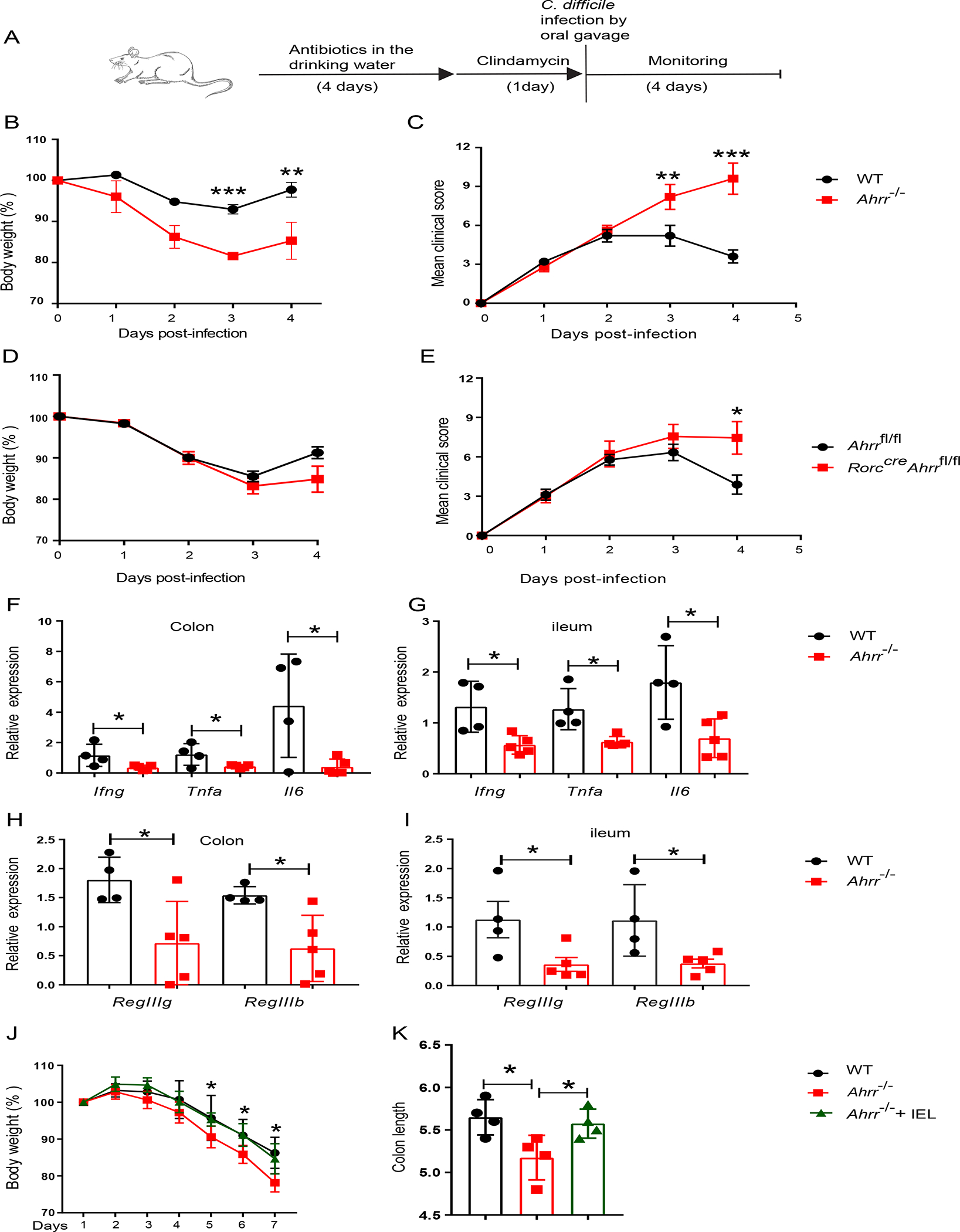
Figure 7. Ahrr deficiency augments susceptibility to intestinal pathology.

(A) Schematic of C. difficile infection in WT and Ahrr−/− mice or Ahrrfl/fl and Rorccre Ahrrfl/fl mice. (B) Body weight and c, clinical score in WT and Ahrr−/− mice. (D) Body weight and (E), clinical score in Ahrrfl/fl and Rorccre Ahrrfl/fl mice. (F-I) Expression of Ifng, Tnfa, Il6, RegIIIg and RegIIIb in colonic and ileal tissues of WT and Ahrr−/− mice upon infection with C. difficile. (J, K) Ahrr−/− mice were reconstituted with WT IEL and, after 3 days, were challenged with 3% DSS for 7 days. (J) % of body weight variation and (K) colon length at day 7 in WT, Ahrr−/− and Ahrr−/− mice reconstituted with WT IEL. Each dot represents an individual mouse. Data are pooled or representative of 2 individual experiments. Statistical significance was determined by Mann-Whitney test. *P<0.05, **P<0.01, ***P<0.001. Please also see Figure S7.
