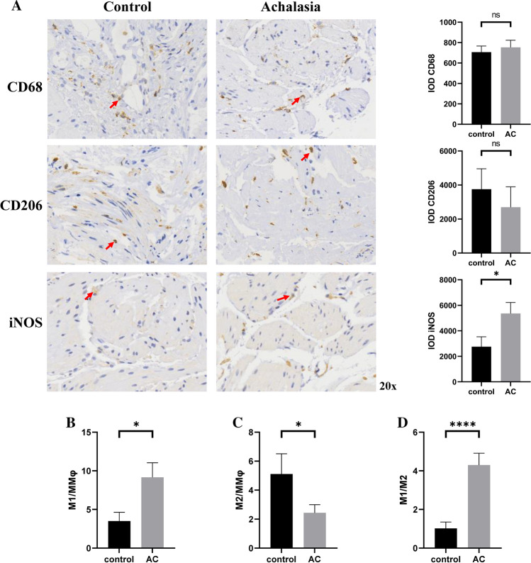
Fig. 2.
Immunohistochemistry and quantification of macrophages in LES tissue samples obtained from patients with achalasia and controls. A The CD68 was used as general marker for macrophages, and antibodies to iNOS were used to identify M1 macrophages and to CD206 to M2 macrophages. Quantification analysis showed that no considerable difference between two groups in total macrophages and M2 macrophages, but level of M1 macrophages in achalasia was higher than that in controls no matter in terms of the absolute number or the proportion of M1 in the total macrophages (B). Statistical differences were also found between two groups in terms of proportion of M2 in the total macrophages (C) and ratio of M1 to M2 (D). iNOS inducible nitric oxide synthase, MMφ muscularis propria macrophages. *P < 0.05; ***P < 0.001. ns not significant

