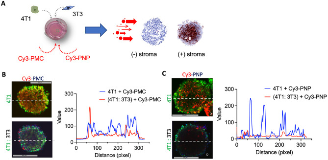
Fig. 4.
Penetration of Cy3-labeled nanoparticles in 3D spheroids using two-photon microscopy. A Schematics showing incubation of Cy3-PMC and Cy3-PNP with 3D 4T1 homospheroids and 4T1:3T3 heterospheroids. 4T1 cells were stained with CellTrackerTMGreen and 3T3 were stained with CellTracker.TMBlue. B, C Two-photon microscopic images show the penetration of Cy3-PMC and Cy3-PNP, respectively and plots with the intensity profile of the red signal across the spheroids (shown with the dotted lines)

