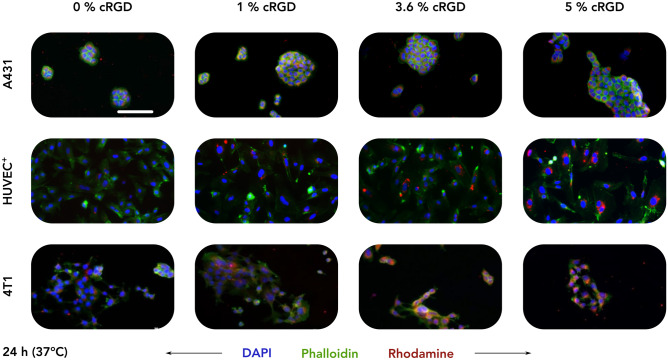
Fig. 2.
Uptake of control and cRGD-decorated CCPM after 24 h of incubation at 37 °C. Representative fluorescence microscopy images depict the uptake of rhodamine-labeled control and cRGD-targeted (1, 3.6, and 5 mol%) CCPM by A431 (human squamous carcinoma), TNFα-activated HUVEC (HUVEC+), and 4T1 (murine triple-negative breast cancer) cells 24 h post incubation, at 37 °C. All four nanoparticle formulations are taken up to a similar extent by A431 cells (αvβ3-integrinnegative cell line), while increasing the cRGD-decoration density results in an enhanced uptake by HUVEC+ and 4T1 cells (αvβ3-integrin.positive cell lines). Color coding: DAPI (nuclei; blue), phalloidin (actin filaments; green), and rhodamine (micelles, red). Scale bar = 100 µm

