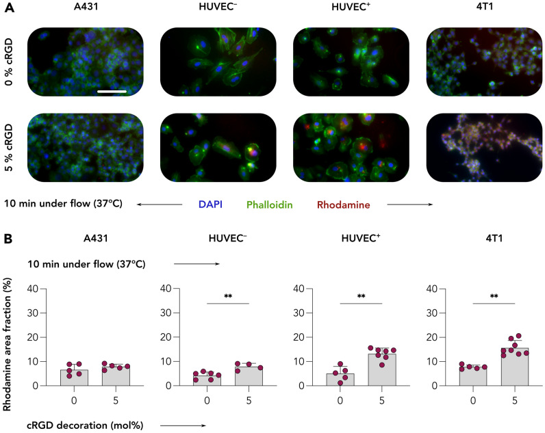
Fig. 5.
Uptake of control and cRGD-decorated CCPM upon 10 min of incubation under flow conditions. A Representative fluorescent microscopy images displaying A431, quiescent (HUVEC−), and TNFα-activated (HUVEC.+) HUVEC, as well as 4T1 cells following incubation with control and 5% cRGD-targeted CCPM under physiological fluid flow for 10 min at 37 °C. Color coding: DAPI (nuclei, blue), phalloidin (actin filaments, green), and rhodamine (CCPM, red). Scale bar = 100 µm. B Quantification of CCPM uptake (i.e., rhodamine area fraction %) shows a threefold increase in the uptake of cRGD-targeted CCPM by ανβ3 integrin-positive cells as compared to control CCPM, exemplifying that cRGD efficiently and rapidly mediates cellular uptake under shear stress conditions. Data are presented as mean ± SD. N = 5–8 biological replicates. Levels of significance were assessed by a Mann–Whitney’s non-parametric two-tailed. p-values: ** < 0.01

