Fig. 6.
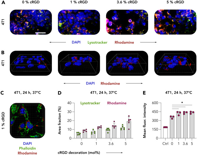
Internalization of cRGD-decorated CCPM. A 4T1 breast cancer cells were incubated with control and cRGD-CCPM for 24 h at 37 °C. LysoTracker was subquentially added to visualize the endosomal-lysosomal compartment. On live-cell imaging, the co-localization (yellow in merged images) of CCPM (red) with endosomes/lysosomes (green) was suggestive for CCPM internalization. Color coding: DAPI (nuclei, blue), LysoTracker (endo/lysosomes, green), and rhodamine (micelles, red). Scale bar = 100 µm. B and C Acquisition of 3D images via two-photon microscopy revealed cRGD-CCPM to be in the perinuclear region, further confirming the cRGD-CCPM internalization by 4T1 cells. Color coding: DAPI (nuclei, blue), phalloidin (actin filaments, green), and rhodamine (micelles, red). D Image analysis shows rhodamine and LysoTracker area fraction % to increase upon the increase of cRGD-decoration density. E Quantification of flow cytometry analysis of micelle binding/uptake by 4T1 cells displays a statistically significant increase in uptake of cRGD-conjugated CCPM in comparison to cRGD-free CCPM. Data are presented as mean ± SD of n = 3 biological replicates; levels of significance were assessed by using a one-way ANOVA followed by Tukey’s correction. p-values: * < 0.05

