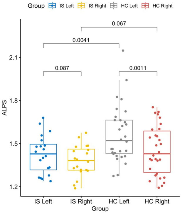
Figure 4.

The graph shows a significantly lower DTI-ALPS index in the left/stroke side of the brain in patients with IS than in HCs (t = −3.02, p = 0.004). The left side was also significantly higher than the right side in HCs (t = 3.62, p = 0.001). Statistics shown in the graph were p-values. DTI-ALPS, diffusion tensor image analysis along the perivascular space; IS, ischemic stroke; HC, healthy controls.
