FIGURE 2.
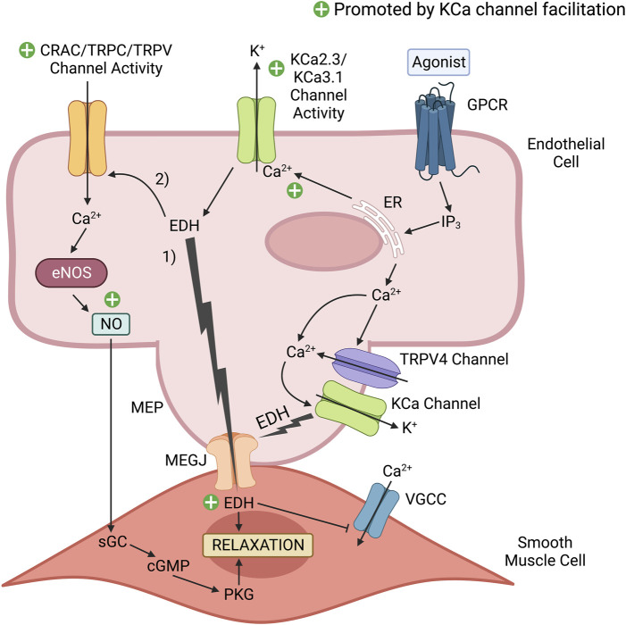
KCa channel positive modulators promote endothelium-dependent vasodilatory signaling. Vasodilatory agonists in the vascular lumen, such as acetylcholine (ACh) bind to their cognate endothelial G-protein coupled receptor (GPCR), which initiates a series of cascading events that results in Ca2+ release from the endoplasmic reticulum (ER). Ca2+ released from the ER can activate endothelial nitric oxide synthase (eNOS) to produce the potent vasodilator nitric oxide (NO). Additionally, the released Ca2+ can activate Ca2+-activated K+ (KCa) channels that induce endothelium-derived hyperpolarization (EDH). 1) This electrical signal transfers to the smooth muscle cells via myoendothelial gap junctions (MEGJs) located within a myoendothelial projection (MEP) to inhibit Ca2+ entry via voltage-gated calcium channels (VGCCs). Reduced Ca2+ entry into the smooth muscle cell limits myosin phosphorylation by Ca2+-dependent myosin light chain kinase (MLCK), and the subsequent level of muscle contraction. 2) In addition to EDH-induced vasorelaxation, EDH increases the electrical driving force to promote external Ca2+ entry into the endothelial cell via Ca2+ permeable channels activated in response to a vasodilatory stimulus (e.g., Ca2+-release activated Ca2+ (CRAC) channels, TRPC3, TRPC4, TRPC6, TRPV1 and TRPV4 channels present in peripheral arteries, such as the aorta). This elevation of Ca2+ in the cytosol and beneath the plasma membrane can promote Ca2+-dependent vasodilatory signaling, such as NO production by eNOS and KCa channel activity. NO readily diffuses to the adjacent smooth muscle to induce relaxation via the cGMP/Protein Kinase G (PKG) pathway. A selective KCa2.X/KCa3.1 positive modulator (e.g., SKA-31) can facilitate the activity of endothelial KCa2.X/KCa3.1 channels, which would enhance EDH and NO production to promote vascular relaxation and healthy endothelial function. The green plus signs highlight mechanisms contributing to endothelium-dependent vasodilation that would be enhanced by KCa channel facilitation. This figure was adapted from CM John et al. (2018) and created with www.BioRender.com.

