Figure 3:

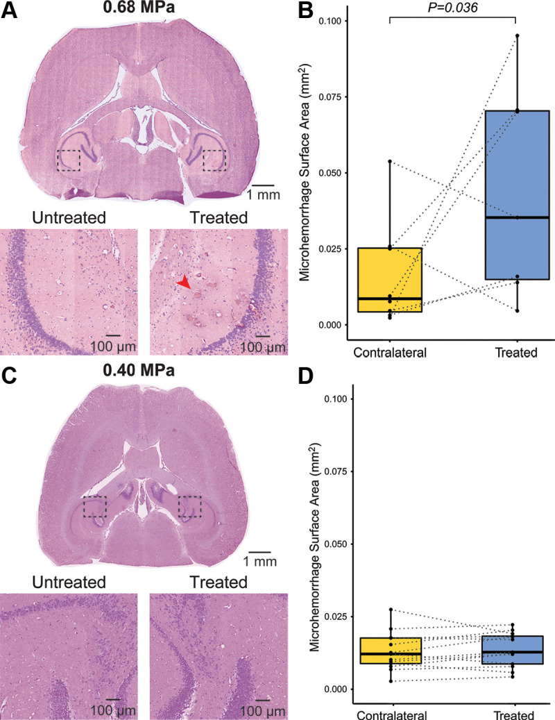
Safety assessment of sonobiopsy. (A) Representative hematoxylin-eosin staining for experiment 1 with 2-month-old mice. (B) There was a significant increase in microhemorrhage surface area (red arrow) in the treated hemisphere (mean, 0.051 mm2 ± 0.039) compared with the untreated hemisphere (mean, 0.016 mm2 ± 0.018; P = .036). (C) Representative hematoxylin-eosin staining for experiment 2 with 6-month-old mice. (D) There was no significant difference in the microhemorrhage density in the treated hemisphere (mean, 0.014 mm2 ± 0.006) compared with the untreated hemisphere (mean, 0.013 mm2 ± 0.007; P = .26; n = 13). Minimal visualized microhemorrhage in the untreated contralateral cerebral hemisphere likely represents artifactual red blood cell extravasation during perfusion, tissue handling, and fixation.
