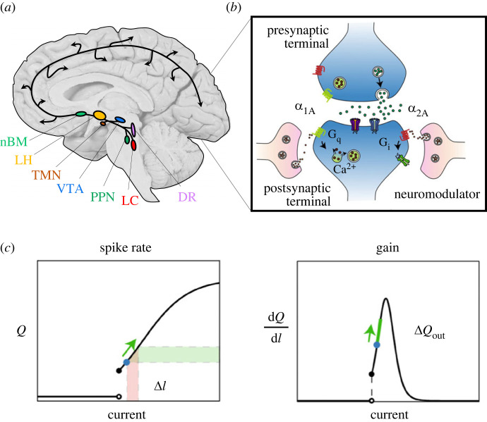
Figure 2.
The AAS alters neuronal gain. (a) The AAS comprises a set of autonomously active nuclei in the brainstem and forebrain that project diffusely around the brain, albeit with idiosyncratic projection patterns. Key: nBM, the cholinergic nucleus basalis of Meynert (nBM; light green); the orexinergic lateral hypothalamus (LH; light orange); the histaminergic tuberomammillary nucleus (TMN; dark orange); the dopaminergic ventral tegmental area (VTA; blue); the cholinergic pedunculopontine nucleus (PPN; dark green); the noradrenergic locus coeruleus (LC; red); and the serotonergic dorsal raphe (DR; purple). (b) Upon release, neuromodulatory ligands predominantly interact with G-protein-coupled receptors (Gq [green] and Gi [green]); (c) this causes a change in the relative spiking output (ΔQout) of a neuron for a given change in current (ΔI), which is also referred to a change in gain (dQ/dI). Key: Q, firing rate. Figure adapted from [21].

