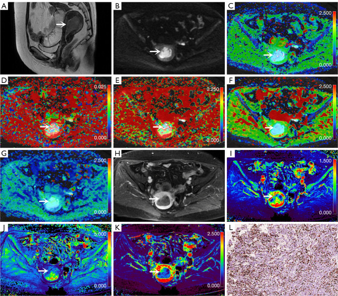
Figure 2.
Images from a 57-year-old woman with endometrial carcinoma (arrows; 5 cm × 2.5 cm × 0.8 cm; Ki-67 =40%; grade 2; stage IA). (A) Sagittal T2-weighted imaging map. (B) Oblique axial diffusion-weighted imaging map (b=800 s/mm2). (C) Oblique axial colored map of the diffusion coefficient, D. (D) Oblique axial colored map of the pseudo diffusion coefficient, D*. (E) Oblique axial pseudo colored map of the perfusion fraction, f. (F) Oblique axial pseudo colored map of the DDC. (G) Oblique axial colored map of the water molecular diffusion heterogeneity index, α. (H) Oblique axial contrast-enhanced map. (I) Oblique axial colored map of the volume transfer constant, Ktrans. (J) Oblique axial colored map of the rate transfer constant, Kep. (K) Oblique axial colored map of the volume of extravascular extracellular space per unit volume of tissue, Ve. (L) Immunohistochemical map of Ki-67 staining (Ki-67 =40%; original magnification 200×). D, diffusion coefficient; D*, pseudo diffusion coefficient; f, perfusion fraction; DDC, distributed diffusion coefficient; α, water molecular diffusion heterogeneity index; Ktrans, volume transfer constant; Ve, volume of extravascular extracellular space per unit volume of tissue; Kep, rate transfer constant.

