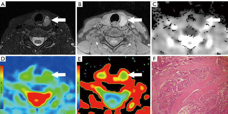
Figure 2.
A 40-year-old female with PTC (arrow) in the left thyroid lobe. (A) Axial T2-WI showed a 16-mm solid nodule with locally irregular margin and a positive taller-than-wide sign: the SIR was 3.13. (B) Axial T1WI: the SIR was 1.35. (C) ADC map; ADC value =0.93×10⁻³ mm2/s. (D) MD map; MD value =1.59×10⁻³ mm2/s. (E) MK map; MK value =0.84. (F) Histopathological HE staining (×100). PTC, papillary thyroid carcinoma; T2WI, T2-weighted imaging; SIR, signal intensity ratio; T1WI, T1-weighted imaging; ADC, apparent diffusion coefficient; MD, mean diffusivity; MK, mean kurtosis; HE, hematoxylin and eosin.

