Abstract
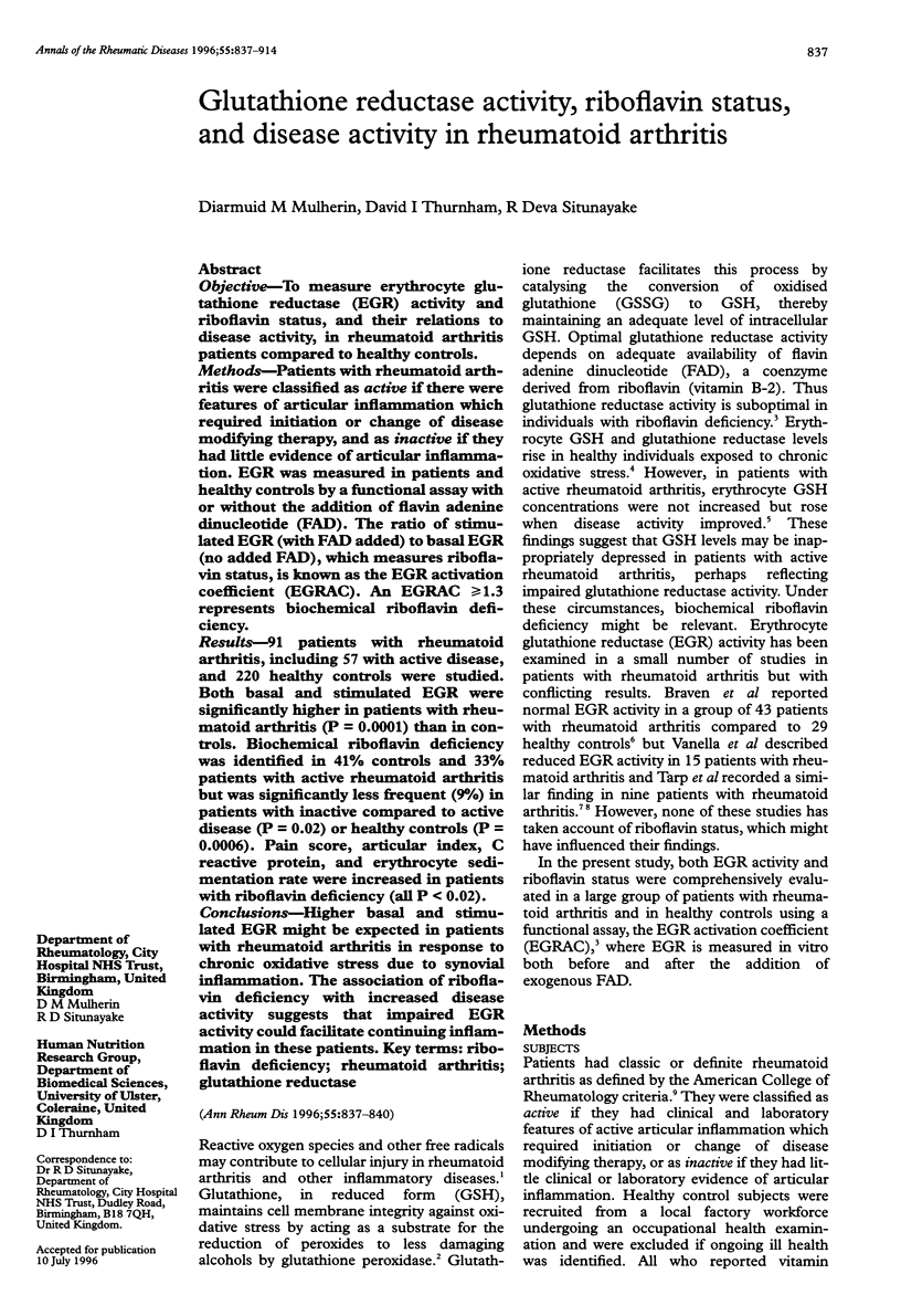
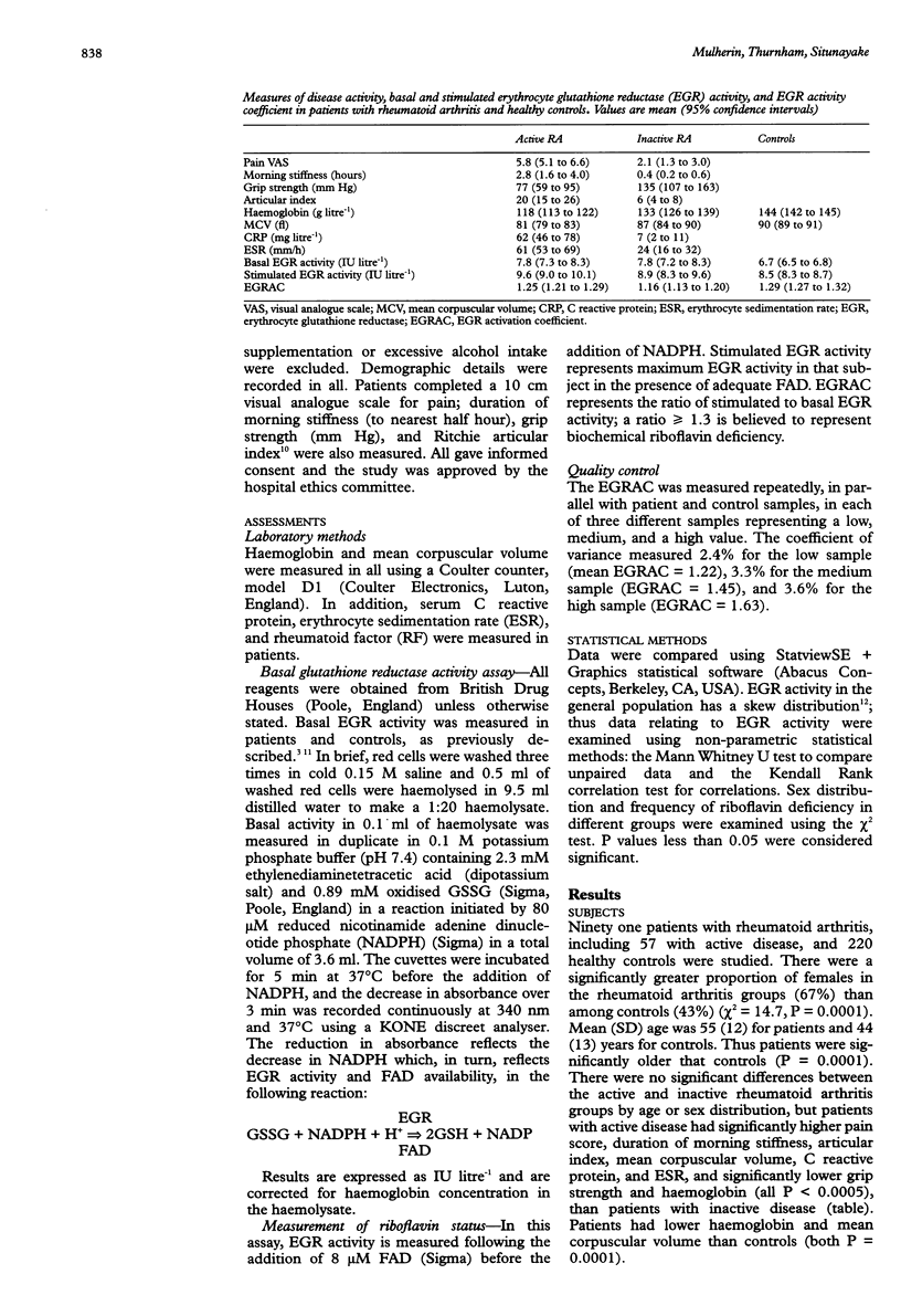
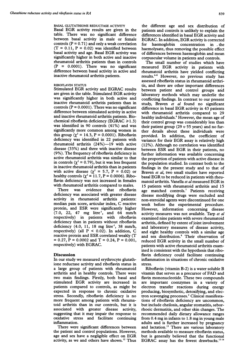
OBJECTIVE: To measure erythrocyte glutathione reductase (EGR) activity and riboflavin status, and their relations to disease activity, in rheumatoid arthritis patients compared to healthy controls. METHODS: Patients with rheumatoid arthritis were classified as active if there were features of articular inflammation which required initiation or change of disease modifying therapy, and as inactive if they had little evidence of articular inflammation. EGR was measured in patients and healthy controls by a functional assay with or without the addition of flavin adenine dinucleotide (FAD). The ratio of stimulated EGR (with FAD added) to basal EGR (no added FAD), which measures riboflavin status, is known as the EGR activation coefficient (EGRAC). An EGRAC > or = 1.3 represents biochemical riboflavin deficiency. RESULTS: 91 patients with rheumatoid arthritis, including 57 with active disease, and 220 healthy controls were studied. Both basal and stimulated EGR were significantly higher in patients with rheumatoid arthritis (P = 0.0001) than in controls. Biochemical riboflavin deficiency was identified in 41% controls and 33% patients with active rheumatoid arthritis but was significantly less frequent (9%) in patients with inactive compared to active disease (P = 0.02) or healthy controls (P = 0.0006). Pain score, articular index, C reactive protein, and erythrocyte sedimentation rate were increased in patients with riboflavin deficiency (all P < 0.02). CONCLUSIONS: Higher basal and stimulated EGR might be expected in patients with rheumatoid arthritis in response to chronic oxidative stress due to synovial inflammation. The association of riboflavin deficiency with increased disease activity suggests that impaired EGR activity could facilitate continuing inflammation in these patients.
Full text
PDF


Selected References
These references are in PubMed. This may not be the complete list of references from this article.
- Adelekan D. A., Thurnham D. I. Effects of combined riboflavin and iron deficiency on the hematological status and tissue iron concentrations of the rat. J Nutr. 1986 Jul;116(7):1257–1265. doi: 10.1093/jn/116.7.1257. [DOI] [PubMed] [Google Scholar]
- Braven J., Ansari N., Figgitt D. P., Fisher A., Luders C., Hickling P., Whittaker M. A comparison of glutathione reductase and glutathione peroxidase activities in patients with rheumatoid arthritis and healthy adults. Br J Rheumatol. 1989 Jun;28(3):212–215. doi: 10.1093/rheumatology/28.3.212. [DOI] [PubMed] [Google Scholar]
- Campbell T. C., Brun T., Chen J. S., Feng Z. L., Parpia B. Questioning riboflavin recommendations on the basis of a survey in China. Am J Clin Nutr. 1990 Mar;51(3):436–445. doi: 10.1093/ajcn/51.3.436. [DOI] [PubMed] [Google Scholar]
- Christensen H. N. Riboflavin can protect tissue from oxidative injury. Nutr Rev. 1993 May;51(5):149–150. doi: 10.1111/j.1753-4887.1993.tb03092.x. [DOI] [PubMed] [Google Scholar]
- Evelo C. T., Palmen N. G., Artur Y., Janssen G. M. Changes in blood glutathione concentrations, and in erythrocyte glutathione reductase and glutathione S-transferase activity after running training and after participation in contests. Eur J Appl Physiol Occup Physiol. 1992;64(4):354–358. doi: 10.1007/BF00636224. [DOI] [PubMed] [Google Scholar]
- Glatzle D., Körner W. F., Christeller S., Wiss O. Method for the detection of a biochemical riboflavin deficiency. Stimulation of NADPH2-dependent glutathione reductase from human erythrocytes by FAD in vitro. Investigations on the vitamin B2 status in healthly people and geriatric patients. Int Z Vitaminforsch. 1970;40(2):166–183. [PubMed] [Google Scholar]
- Munthe E., Kåss E., Jellum E. D-penicillamine-induced increase in intracellular glutathione correlating to clinical response in rheumatoid arthritis. J Rheumatol Suppl. 1981 Jan-Feb;7:14–19. [PubMed] [Google Scholar]
- ROPES M. W., BENNETT G. A., COBB S., JACOX R., JESSAR R. A. 1958 Revision of diagnostic criteria for rheumatoid arthritis. Bull Rheum Dis. 1958 Dec;9(4):175–176. [PubMed] [Google Scholar]
- Ritchie D. M., Boyle J. A., McInnes J. M., Jasani M. K., Dalakos T. G., Grieveson P., Buchanan W. W. Clinical studies with an articular index for the assessment of joint tenderness in patients with rheumatoid arthritis. Q J Med. 1968 Jul;37(147):393–406. [PubMed] [Google Scholar]
- Salkie M. L., Simpson E. The erythrocyte glutathione reductase activity (E.C. 1.6.4.2) of healthy blood donors. J Clin Pathol. 1970 Nov;23(8):708–710. doi: 10.1136/jcp.23.8.708. [DOI] [PMC free article] [PubMed] [Google Scholar]
- Stefanovic-Racic M., Stadler J., Evans C. H. Nitric oxide and arthritis. Arthritis Rheum. 1993 Aug;36(8):1036–1044. doi: 10.1002/art.1780360803. [DOI] [PubMed] [Google Scholar]
- Tarp U., Stengaard-Pedersen K., Hansen J. C., Thorling E. B. Glutathione redox cycle enzymes and selenium in severe rheumatoid arthritis: lack of antioxidative response to selenium supplementation in polymorphonuclear leucocytes. Ann Rheum Dis. 1992 Sep;51(9):1044–1049. doi: 10.1136/ard.51.9.1044. [DOI] [PMC free article] [PubMed] [Google Scholar]


