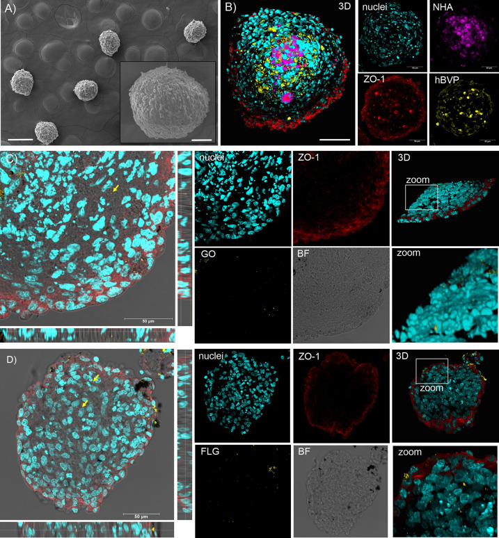
Figure 4.
GO and FLG interactions with a 3D human multicellular assembloid model of BBB: SEM and confocal microscopy analysis. (A) SEM micrographs of hMCA showing their spherical morphology. (B) Confocal imaging and 3D reconstruction of hMCA: prestained NHA and hBVP are shown in purple and yellow, respectively; ZO-1 stained hCMEC/D3 tight junctions are shown in red. Representative confocal XY planes, Z projections, and 3D reconstructions from a 20 μm hMCA slice incubated with 10 μg/mL of GO (C) or FLG (D) for 24 h. Nuclei (Hoechst staining) are visualized in cyan, the two GRMs observed through LR mode are reported in yellow, and ZO-1 immunoreactivity is shown in red.

