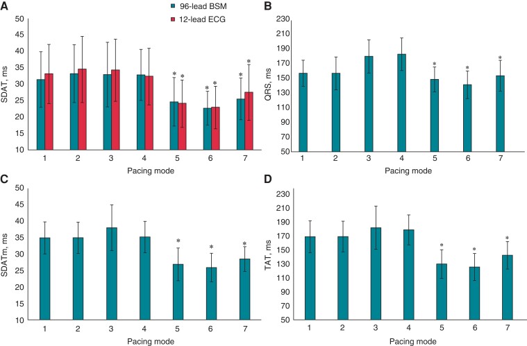
Figure 3.
Measures of electrical dyssynchrony in different pacing modes. (A) Standard deviation of activation time measured from 96-lead mapping system (96-lead SDAT) and from 12-lead ECG; (B) mean QRS duration calculated from standard limb leads; (C) SDATm; and (D) TAT determined from reconstructed myocardial activation times. Pacing modes: 1, Sinus rhythm; 2, atrial pacing; 3, sequential LV pacing (AV delay 120 ms); 4, sequential RV pacing (AV delay 120 ms); 5, sequential biventricular pacing (AV delay 120 ms, VV delay 0 ms); 6, sequential biventricular pacing (AV delay −20 ms intrinsic PQ, VV delay 0 ms); 7, sequential biventricular pacing (AV delay −20 ms intrinsic PQ, VV delay LV −40 ms over RV). *P < 0.01 in comparison with Pacing Mode 1. SDAT, standard deviation of activation time; SDATm, standard deviation of reconstructed myocardial activation time; TAT, total activation time.

