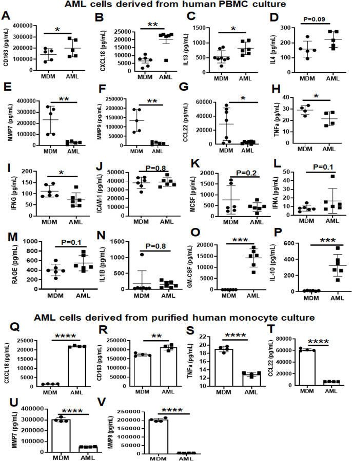
Fig 7. AML cells release several inflammation-related proteins.
(A-I) PBMCs were exposed to ALL cocktail [surfactant (Infasurf) and cytokines (GM-CSF, TGF-β, IL-10)] for 6 days on alternative days or left untreated (MDM). Cell supernatants were collected and release of several inflammation-related proteins was analyzed simultaneously by Luminex technology. Like HAM, AML cells released increased levels of (A) CD163, (B) CXCL18, (C) IL13 and (D) IL4, and decreased levels of (E) MMP7, (F) MMP9, (G) CCL22, (H) TNFα and (I) IFNG compared to MDM. AML cells and MDM released similar quantities of soluble (J) ICAM-1, (K) MCSF, (L) IFNA, (M) RAGE and (N) IL1B, and there was significantly more (O) GM-CSF and (P) IL-10 in the supernatants collected from AML cells than from MDM. Data are mean ± SEM; each dot indicates results from 1 donor (n=5–8) and analyzed by Unpaired Student’s ‘t’ test. *p≤0.05, ** p≤0.01, *** p≤0.001. (Q-V) Monocytes were purified by EasySep™ human monocyte isolation kit by negative selection magnetic sorting of healthy human PBMCs on Day 0 and exposed to ALL cocktail treatment [surfactant (Infasurf) and cytokines (GM-CSF, TGF-β, IL-10)] for 6 days on alternative days or left untreated (MDM). Cell supernatants were collected and release of inflammation-related proteins was analyzed simultaneously by Luminex technology. AML cells differentiated from isolated monocytes release higher (Q) CXCL18 and (R) CD163, and lower (S) TNFα, (T) CCL22, (U) MMP7 and (V) MMP9 amounts than MDM, similar to those cells differentiated from PBMCs. Data are expressed as mean ± SEM; each dot indicates results from 1 donor (n=4) and analysed by Unpaired Student’s ‘t’ test. ** p≤0.01, ****p≤0.0001.

