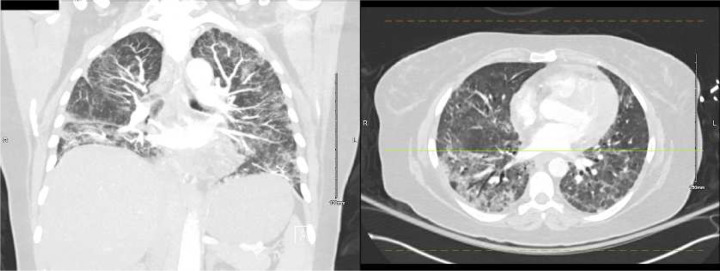
Figure 5.
Outpatient chest computed tomography two days before the patient’s third admission to the hospital, showing ground glass and airspace opacities in the lower lungs differening in distribution from the first admission, with a few focal areas of honeycombing, most prominently in the left upper lobe.

