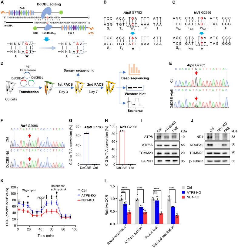
Fig. 1. DdCBE-mediated ATP8/ND1 KO in rat C6 cells.
(A) Strategy of KO mtProteins by introducing the stop codon using DdCBE. (B and C) The DdCBE-mediated C·G-to-T·A conversion at Atp8 G7783 (B) and Nd1 G2996 (C). (D) The work flow for DdCBE-mediated mtProteins KO in C6 cells. (E and F) Sanger sequencing chromatogram of spacer for Atp8 G7783 site (E) and Nd1 G2996 site (F) at day 3 after the first-round enrichment [first fluorescence-activated cell sorting (FACS)] of double-positive C6 cells. (G and H) The frequency of the DdCBE-mediated C·G-to-T·A conversion at Atp8 G7783 (G) and Nd1 G2996 (H) sites was analyzed using deep sequencing at day 7 after the second-round enrichment (second FACS). n = 3 technical replicates. Data were presented as means ± SD. Untreated C6 cells were used as control (Ctrl). (I and J) The protein levels of ATP8 (I) and ND1 (J) were detected by Western blot in rat C6 cells treated with DdCBE or flunarizine (FNZ). (K) Mitochondrial respiratory capacity was indicated by the oxygen consumption rate (OCR) in Ctrl and ATP8-/ND1-KO C6 cells. (L) Relative values of OXPHOS parameters from (K). n = 5 for technical replicates. Data were presented as means ± SD. ns, not significant; ***P ≤ 0.001 and ****P ≤ 0.0001, by one-way analysis of variance (ANOVA) test paired with a Tukey’s post hoc test.

