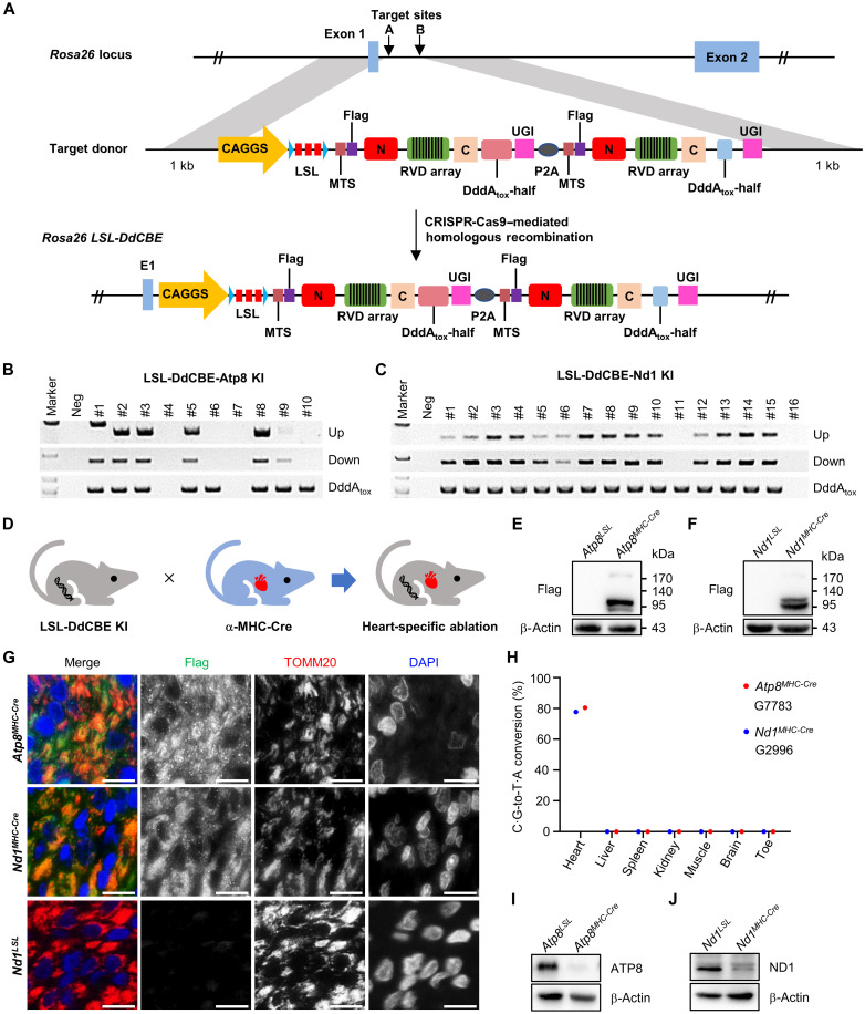
Fig. 2. DdCBE-mediated ATP8/ND1 cKO in rats.
(A) The incorporation of LSL-DdCBE into rat Rosa26 locus using CRISPR-Cas9–mediated homologous recombination. (B and C) Genotyping of LSL-DdCBE-Atp8 KI (B) and LSL-DdCBE-Nd1 KI (C) F0 rats by PCR with upstream primer pair (Up band), downstream primer pair (Down band), and internal primer pair (DddAtox band). (D) The mtProteins KO in the heart tissue induced by crossing LSL-DdCBE KI rat with α-MHC-Cre rat. (E and F) The DdCBE expression in the heart tissue of Atp8MHC-Cre (E) and Nd1MHC-Cre (F) rat was detected by Western blot using the anti-Flag antibody. (G) The localization of DdCBE in mitochondrial of Atp8MHC-Cre and Nd1MHC-Cre heart tissues. The anti-Flag (green) for DdCBE, anti-TOMM20 for mitochondria (red), and 4′,6-diamidino-2-phenylindole (DAPI) for nucleus (blue). Scale bars, 10 μm. (H) The frequency of DdCBE-mediated C·G-to-T·A conversion at Atp8 G7783 and Nd1 G2996 sites in the heart, liver, spleen, kidney, muscle, brain, and toe of Atp8MHC-Cre and Nd1MHC-Cre rats was analyzed by deep sequencing. (I and J) Protein level of ATP8 (I) and ND1 (J) in tissues of Atp8MHC-Cre and Nd1MHC-Cre was detected by Western blot, respectively.

