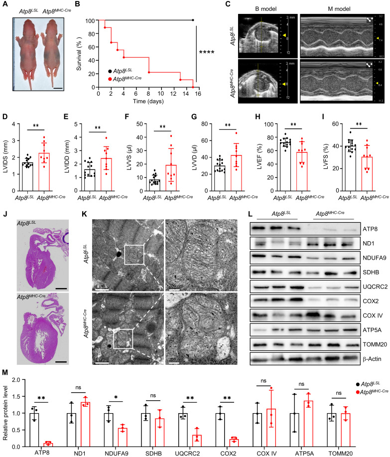
Fig. 3. Heart-specific depletion of ATP8 causes heart failure in rats.
(A) The whole-body image of 3-day-old Atp8LSL and Atp8MHC-Cre pups. Scale bars, 1 cm. (B) The survival curve of Atp8LSL and Atp8MHC-Cre rats. n = 10 biological replicates for each group. ****P ≤ 0.0001 by log-rank test. (C) The snapshot of echocardiography for Atp8LSL and Atp8MHC-Cre rat. (D to I) The cardiac structure and function parameters of Atp8LSL and Atp8MHC-Cre rats calculated from echocardiography in (C). LVIDS (D), LVIDD (E), LVVS (F), (LVVD) (G), LVEF (H), and LVFS (I). n = 14 for the Atp8LSL group and n = 9 for the Atp8MHC-Cre group. Data were presented as means ± SD. **P ≤ 0.01 by Student’s unpaired two-tailed t test. (J) The H&E image of heart tissue from Atp8LSL and Atp8MHC-Cre rats. Scale bars, 1 mm. (K) The TEM image of mitochondria in heart tissues of Atp8LSL and Atp8MHC-Cre rats. (L) The mitochondrial protein level was detected by Western blot in heart tissues of Atp8LSL and Atp8MHC-Cre rats. (M) Relative protein level of mitochondrial proteins from (L). n = 3 biological replicates for each group. Data were presented as means ± SD. *P ≤ 0.05 and **P ≤ 0.01 by Student’s unpaired two-tailed t test. ns, not significant.

